

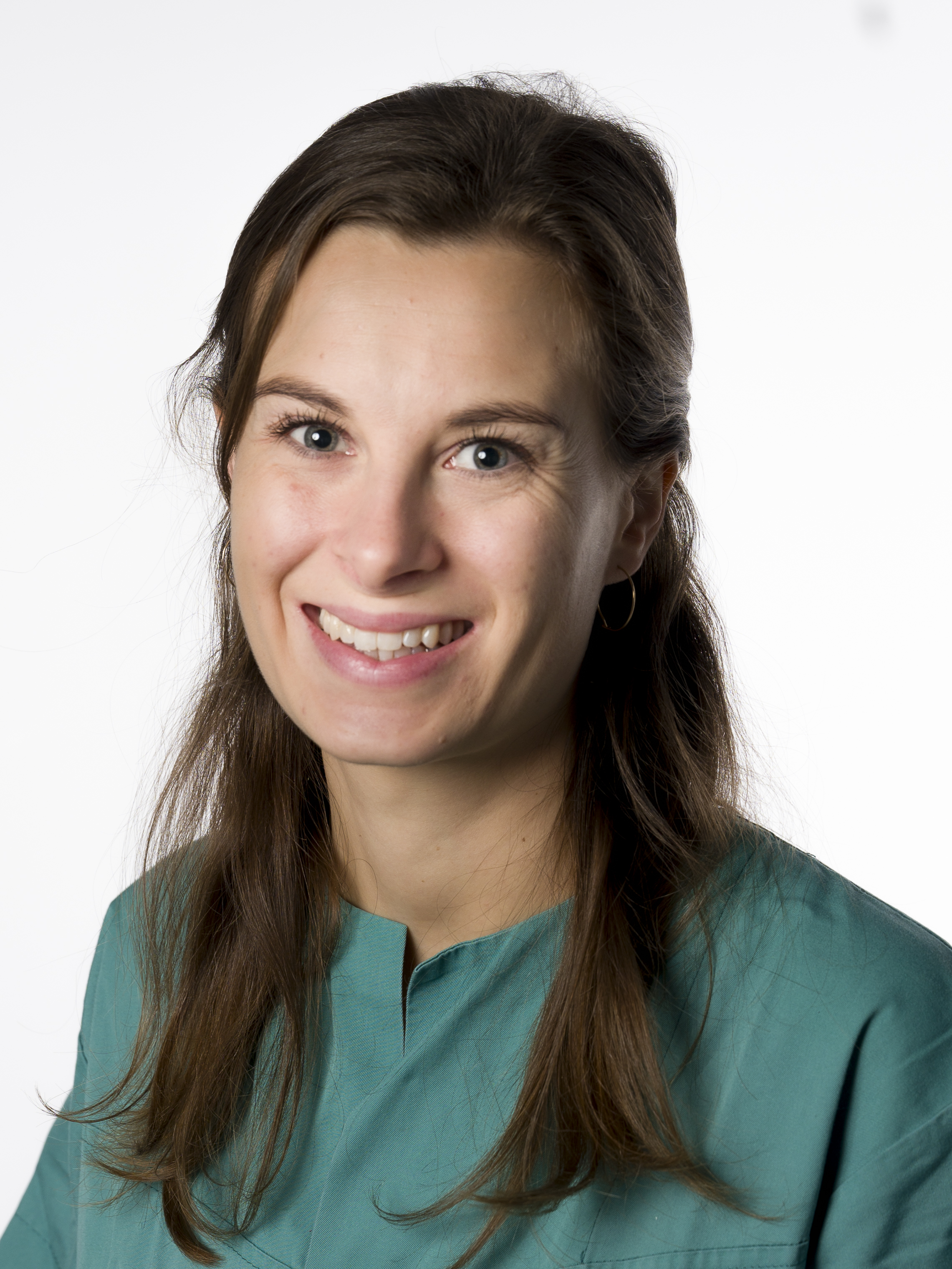
Celine Austrheim Krefting is a medical engineer at Oslo University Hospital, as demand for personalised care grows, hospitals are increasingly adopting point-of-care 3D labs to bring these capabilities in-house. Celine and her team built an in-house lab at Oslo University Hospital back in 2021.
OPN: What drove you to choose a career in medical engineering?
CAK: I have always been interested in both the medical, and engineering fields. When I discovered that I had the opportunity to study a combination of the two, the choice was easy. After completing my master’s degree, I was fortunate to secure a position as a medical engineer, and since then I have held two different roles within the field. I have been happy in both roles, which is why I have chosen to continue my career in medical engineering.
OPN: Could you tell us more about your work as an engineer, making 3D models based on CT and MRI scans?
CAK: My work is divided between administrative tasks, such as developing a security quality management system, and more creative tasks, like producing anatomical 3D models and surgical guides. What is quite unique about working at a hospital, is that my team work closely with surgeons. We are able to join outpatient consultations as well as observe surgeries. This is valuable for our professional development and is essential for effective learning.
In short, our workflow for creating 3D models starts with receiving patient cases from the surgeons, requesting either an anatomical model or a full digital surgery with surgical guides. The request come through our secure patient system. The engineers have learnt enough anatomy to independently create a 1:1 3D model from CT, MRI or ultrasound images, a process called segmentation. In this step we identify and outline the anatomical structures requested by the doctors. When outlining, our software creates 3D models in a semi-automatic process. Once the model is complete, it must be quality-checked together with the surgeons before printing.
If the surgeon requests a digital surgery planning and/or a surgical guide, we will meet with them to plan how the procedure will be performed. Engineers can do a lot of the work on our own, but we typically need at least two to three collaborative meetings to determine for example where cuts, drill holes and fixation plates should be placed.
OPN: How could the models help support surgeons and their patients during orthopaedic surgery?
CAK: The anatomical models are very valuable for surgeons during preoperative planning. They help improve understanding the patient’s condition, support discussions during outpatient consultations, and can even be used to simulate parts of the surgery in advance. Many of our surgeons also use the models for teaching or as visual aids during presentations at for example conferences.
Research has shown that digital planning and surgical guides can reduce operating time, radiation exposure and blood loss. Our internal research indicate that 3D-printing enhances surgical understanding, increase surgical precision and provides strong support for validating surgical concepts. We also see that having an in-house 3D-lab is both time- and cost-effective for the hospital, in addition to strengthening interdisciplinary collaboration.
OPN: What is planned for the next stage of your research and engineering development?
CAK: We have already started to expand our expertise in designing surgical guides within hand surgery and cranio-maxillofacial surgery, which are well-established applications at many other point-of-cares. These are interesting and large fields that we would like to work more with, we just simply haven’t had the time until now.
We will also further explore the use of augmented/ virtual reality in cases where 3D printing does not